
In the medical field, many types of diagnostic imaging techniques are used to identify a medical condition in patients. Diagnostic imaging is a tool that doctors can utilize to “look inside” patients and there are many methods used to achieve this. Everything from ultrasounds to x-rays are used to produce 2D and 3D images that can be reviewed by medical professionals to make critical diagnoses. The imaging techniques that use radiation such as x-rays, radio frequencies, and gamma rays often require shielding to protect patients and healthcare workers as well as provide clear and accurate images.

Radiography
Radiography uses ionizing radiation like gamma or x-rays to produce an image. X-Ray is a conventional and commonly used form of radiography that produces 2D images by projecting the image of a patient on a film or detector that produces a computer image. The ionizing radiation is stopped by some tissues or materials (like bones or metal) but passes through or partially through soft tissue. An X-Ray image then is most useful when looking at bones in the body and is often the first method of imaging due to it being inexpensive and quick procedure.
Another type of Radiography is Computed Tomography, which takes images of the body from multiple angles to produce multiple 2D images of the body. These 2D images can be combined to create 3D images of the body.
Nuclear Imaging
Nuclear imaging typically uses a short-lived radioisotope tracer injected into the body. Commonly used medical tracers are iodine, ![]() technetium-99 (decayed from molybdenum-99), gallium-67 and many others. As a tracer decay in the body, gamma radiation is released and then detected by a gamma scanner. The information from the gamma scanner can then be put together to form an image. Scintigraphy, SPECT, and Proton emission tomography (PET) are all examples of imaging techniques that make use of Nuclear Medicine tracers. Nuclear imaging differs from other imaging methods as the radiation occurs from inside the body rather than from a radiation source outside the body.
technetium-99 (decayed from molybdenum-99), gallium-67 and many others. As a tracer decay in the body, gamma radiation is released and then detected by a gamma scanner. The information from the gamma scanner can then be put together to form an image. Scintigraphy, SPECT, and Proton emission tomography (PET) are all examples of imaging techniques that make use of Nuclear Medicine tracers. Nuclear imaging differs from other imaging methods as the radiation occurs from inside the body rather than from a radiation source outside the body.
Both nuclear imaging and radiography make use of lead shielding. Nuclear medicine tracers need to be shielded from the manufacturing stage to final use in the patient. Often nuclear medicine makes use of shielding products like tungsten and lead pigs, lead lined cabinets, shielded carts, and storage containers. Radiography requires lead or lead alternative shielding to shield from backscatter radiation and to prevent adjacent rooms or areas next to the imaging rooms from long term exposure. Sheet lead products like MarShield E-Z Shielding Panels can be very useful to provide radiation protection. Lead lined mobile barriers can also be used to provide additional shielding where needed and windows can be protected by using lead glass or acrylic.
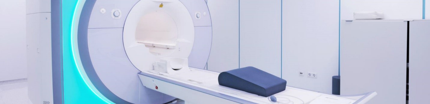
Magnetic Resonance Imaging (MRI)
MRIs produce images by interacting with hydrogen nuclei of water molecules in the human body. The hydrogen nuclei align in the MRI’s magnetic field. The MRI then produces a Radio Frequency (RF) pulse that disrupts the direction of the hydrogen. When the pulse is turned off, the atoms realign with the magnetic field and the realignment produces radio waves that can be detected, and an image can be constructed out of these signals.
Because MRI machines do not use Ionizing radiation, they are generally considered to be a safer option for medical imaging. The hazards related to MRI machines are related to the large magnetic field produced by the magnet inside the machine. When operational, metal objects will be pulled towards the machine so all magnetic materials must be removed from the patient and room. During operation, if a barrier is needed to shield from nuclear medicine tracers, it must be constructed completely from nonmagnetic material.
An MRI machine does require shielding, but its purpose is to produce as clear an image as possible. Noise RF waves can easily interfere with the image because the MRI relies on RF signals being generated by hydrogen nuclei realigning with the MRI’s magnetic field. RF waves are present all around us from radio waves to cell phone signals and wireless internet. To shield outside RF waves, the room is enclosed with conductive material and electrically grounded. This is also commonly known as a Faraday Cage. MarShield’s RF Shielding system typically uses either copper or galvanized steel panels. The copper panels can be soldered together or in the case of the galvanized steel, joined by steel framing. The doors, windows, vents, plumbing, and electrical are all specially designed to shield from RF Radiation.
Medical Diagnostic Imaging uses a wide variety of techniques. Knowing which produces radiation that requires shielding is important to properly protect patients and healthcare workers on the job, as well in some cases providing clearer images. MarShield has provided shielding, protection, and storage solutions for medical imaging from X-Rays to MRI and more.
If you would like to learn more about MarShield’s shielding capabilities in the imaging industry, you can visit the MarShield website or you can contact MarShield directly, and we will help you find the best radiation shielding solution for you.